Transposition of the Great Arteries

What is it?
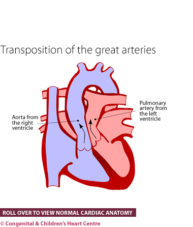
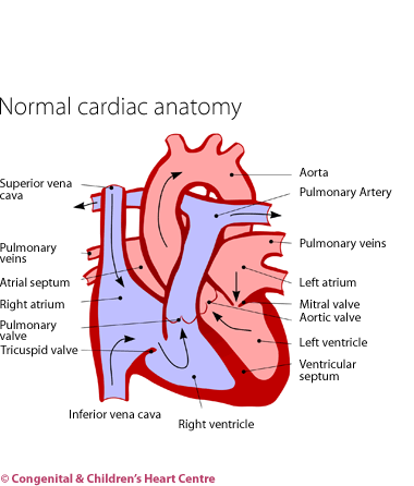
Transposition of the great arteries (or TGA) is a condition whereby the two arteries from the heart are swapped, so that they end up the wrong way around. In other words, the main pumping chamber to the body (left ventricle) is connected to the blood vessel to the lungs (pulmonary artery) and the pumping chamber to the lungs (right ventricle) is connected to the blood vessel to the body (aorta), instead of vice versa.
This means that blue blood returning from the body, which should go to the lungs to receive oxygen and become pink, actually goes straight back to the body. Blood that has just returned from the lungs and is pink and which should get pumped to the body goes straight back to the lungs. This situation is obviously not compatible with life unless there is some mixing of these two circulations. This occurs at two places.
First, in every unborn baby there is a hole (patent foramen ovale or atrial septal defect) between the two collecting chambers (atria) of the heart. In a child with TGA, this allows some mixing to occur. Second, in the circulation of the unborn child, there is a pipe called the ductus arteriosus, which allows blood to pass from the lung circulation into the body circulation. This also allows mixing of blood in the transposition circulation. After birth, the hole and the duct tend to shut off. Keeping them open is the first step in the treatment of this condition.
How many people get it?
It has been estimated that there are 4 cases of TGA out of every 10,000 live births accounting for about 4% of all congenital heart disease.
Who gets it?
Anyone can get transposition of the great arteries. Whatever its cause, it is present from birth and probably from very early on in the pregnancy (possibly the 7th week after conception).
What are the signs and symptoms?
The baby is usually well after birth but becomes progressively blue on the lips and tongue over the course of the first day of life. At first the baby is usually fairly well and not breathless. If the blueness is not spotted promptly, the baby becomes progressively unwell.
With the advent of fetal echocardiography (ultrasound scanning of the unborn child's heart), the diagnosis can sometimes be made before the child is born. Arrangements can be made for the infant to be born at or near a cardiac centre.
What kind of tests might I have?
Infants are usually referred to a paediatric cardiologist, who will investigate further. After questioning the parents to obtain the child's history and examining the child, several tests will be organised.
These will usually consist of an electrocardiogram (ECG) to measure the electrical activity of the heart and a chest X-ray to visualise the heart and lungs.
The diagnosis is made by echocardiography. This ultrasound scan will show that the vessels to the body and lungs are the wrong way around. Some blood tests may also be necessary.
What is the treatment?
- Infusion of a medicine called prostaglandin to keep the duct open and allow mixing of the two circulations
- Balloon atrial septostomy at cardiac catheterisation. This is performed under sedation or light anaesthesia. A catheter (tube) is inserted into the leg vein or umbilicus (tummy button) and a balloon introduced into the heart. On withdrawal this makes a hole (atrial septal defect) between the top 2 chambers of the heart (atria) to allow increased mixing of the blood.
- Surgical repair - the 'switch procedure'. The arteries are cut and swapped into the correct position. The most difficult part of the operation is to transfer the coronary arteries across as well.
What is the prognosis?
The prognosis of TGA is generally good, with survival without complications nearly 100%. In the long term, these children fare well and most are completely without symptoms and lead normal lives.
Download Transposition of the Great Arteries PDF![]()
Further information at The Children's Heart Federation

