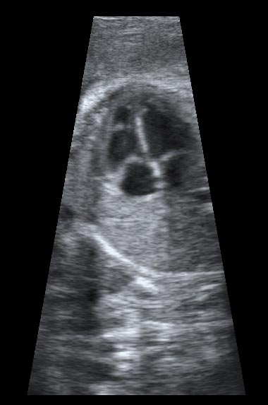
Fetal echocardiography
What is it?
This is a ultrasound scan performed on pregnant women by fetal cardiologists or technicians to provide views of the heart of the unborn child. High frequency sound, too high to hear, is beamed through the mother's abdomen to the fetal heart and the reflection is viewed as an image on the screen, rather like sonar in submarines. As well as structural information about the heart, the use of a technique called Doppler allows the speed and direction of blood in the heart to be ascertained. This can be shown as a coloured image on the screen or as a waveform.
Fetal echocardiography is performed on two groups of pregnant women. Firstly, those in whom the anomaly scan showed a suspected abnormality of the fetal heart
Secondly, those in whom the routine anomaly scan was normal but whose babies are at 'high risk' of heart problems. This may because there is
- Strong family history of cardiac disease
- Increased nuchal thickness detected during the nuchal scan at 12 to 14 weeks of pregnancy
- Presence of abnormalities of the chromosomes following amniocentesis or chorionic villous sampling (CVS)
- Presence of Down or other syndromes in the fetus
- Diseases in the pregnant woman such as diabetes or lupus
- Use of drugs during pregnancy
If a cardiac defect is detected, the fetal consultant will discuss the findings with the patient. This will begin with an explanation of the abnormality found and proceed to an evaluation of the likely treatment after birth. All available options would be discussed.
A repeat scan may be necessary to assess any progression of the condition. Plans would be made to ensure that the baby was born at or near a centre that could care for a newborn with heart problems. After birth, the infant would be transferred to the cardiac centre for reassessment and treatment.
What is going to happen?
This is identical to other ultrasound scans in pregnancy. While lying down in a darkened room, special ultrasound water-based gel is placed on the echo transducer (probe) and the pregnant woman's abdomen. The transducer is then placed at various points on the abdomen and very light pressure applied to obtain the best image possible.
The partner, or alternatively, a friend or relative of the woman may be present for the investigation. An explanation of the findings is given at the end of the scan.
How do I feel during the procedure?
Other than gel on the abdomen and light pressure from the transducer, there is no sensation. The ultrasound itself cannot be felt or heard.
How long does it take?
The scan will last from 10 to 60 minutes, depending on the difficulty in obtaining good views or the complexity of the heart defect.
Is there anything I should or should not do?
There is nothing specific that you should or should not do.
Are there any side effects?
No conclusive adverse effects have been proven. Millions of ultrasound scans have been performed worldwide.

